Abstract
Early events, such as formation of the cell wall, first nuclear division and first unequal division of the zygote, were examined following in vitro fusion of single egg and sperm protoplasts of maize ( Zea mays L.). The time course of these events was determined. The formation of cell wall components was observed 30 sec following egg—sperm fusion and proceeded continuously thereafter. Within 15 h after fusion most of the organelles became more densely grouped around the nucleus of the zygote. In the in vitro produced zygote the location of the cell organelles and of the dividing nucleus showed polarity. Two nucleoli were first observed 18 h after gamete fusion. The zygotic nucleus remained undivided for about 40 h. The first cell division was observed 40–60 h, generally 42–46 h, after egg—sperm fusion. The non‐fused egg cell could be triggered to sporophytic development in vitro by pulses of high amounts of 2,4‐D. Without such a treatment, cultured egg cells of different maize lines did not divide. Although nuclear fusion seemed to occur, fusion products of two egg cells also did not divide. Cell wall formation was incomplete and non‐uniform, showing a polarity of cultured egg cells and fusion products of two egg protoplasts. Cell division was also induced after fusion of maize egg with sperms of genetically remote species, such as Coix, Sorghum, Hordeum or Triticum . These gametic heterologous fusion products developed to microcalli. Moreover, cell division occurred in fusion products of an egg and a diploid somatic cell‐suspension protoplast from maize.
Abstract from the paper’s OpenAlex record, shown for indexing and research. Copyright remains with the authors or the publisher. Read at the source ↗
The text the model read, with the source record’s inline markup (italics, sub- and superscripts) rendered and its character entities decoded; quote offsets refer to the raw text.
Claims 11
Claim · model-written summary
Maize zygotes began forming cell wall components 30 sec after egg–sperm fusion, continuing thereafter.
 Zea mays
Organisms pictured · Representative pictures of the matched species, not figures from the paper.

Verbatim
e0“The formation of cell wall components was observed 30 sec following egg—sperm fusion and proceeded continuously thereafter.”
e1“protoplasts of maize ( Zea mays L.)”
Claim · model-written summary
Most organelles grouped more densely around the maize zygote nucleus within 15 h of fusion.
Organisms pictured · Representative pictures of the matched species, not figures from the paper.
Verbatim
e0“Within 15 h after fusion most of the organelles became more densely grouped around the nucleus of the zygote.”
e1“protoplasts of maize ( Zea mays L.)”
Claim · model-written summary
The organelles and dividing nucleus in the in vitro maize zygote showed polarity.
- Organ / stage
- zygotee0
- Outcome
- location of the cell organelles and of the dividing nucleus showed polaritye0
Organisms pictured · Representative pictures of the matched species, not figures from the paper.
Verbatim
e0“In the in vitro produced zygote the location of the cell organelles and of the dividing nucleus showed polarity.”
e1“protoplasts of maize ( Zea mays L.)”
Claim · model-written summary
Two nucleoli following maize gamete fusion.
Organisms pictured · Representative pictures of the matched species, not figures from the paper.
Verbatim
e0“Two nucleoli were first observed 18 h after gamete fusion.”
e1“protoplasts of maize ( Zea mays L.)”
Claim · model-written summary
zygotic nucleus remained undivided following maize gamete fusion.
Organisms pictured · Representative pictures of the matched species, not figures from the paper.
Verbatim
e0“The zygotic nucleus remained undivided for about 40 h.”
e1“protoplasts of maize ( Zea mays L.)”
Claim · model-written summary
first cell division following maize gamete fusion.
Organisms pictured · Representative pictures of the matched species, not figures from the paper.
Verbatim
e0“The first cell division was observed 40–60 h, generally 42–46 h, after egg—sperm fusion.”
e1“protoplasts of maize ( Zea mays L.)”
Claim · model-written summary
Pulses of high amounts of 2,4-D induced sporophytic development in unfused maize egg cells, unlike untreated cells.
- Organ / stage
- egg celle0
- Treatment
- pulses of high amounts of 2,4‐De0
- Outcome
- sporophytic developmente0
- Comparator
- Without such a treatmente0
Not reportedDirection
Organisms pictured · Representative pictures of the matched species, not figures from the paper.
Verbatim
e0“The non‐fused egg cell could be triggered to sporophytic development in vitro by pulses of high amounts of 2,4‐D. Without such a treatment, cultured egg cells of different maize lines did not divide.”
Claim · model-written summary
Fusion products of two maize egg cells did not divide despite apparent nuclear fusion.
- Outcome
- fusion products of two egg cells also did not dividee0
Organisms pictured · Representative pictures of the matched species, not figures from the paper.
Verbatim
e0“Although nuclear fusion seemed to occur, fusion products of two egg cells also did not divide.”
e1“protoplasts of maize ( Zea mays L.)”
Claim · model-written summary
Cultured maize egg cells and two-egg fusion products showed incomplete, nonuniform, polar cell wall formation.
- Outcome
- Cell wall formation was incomplete and non‐uniforme0
Organisms pictured · Representative pictures of the matched species, not figures from the paper.
Verbatim
e0“Cell wall formation was incomplete and non‐uniform, showing a polarity of cultured egg cells and fusion products of two egg protoplasts.”
e1“protoplasts of maize ( Zea mays L.)”
Claim · model-written summary
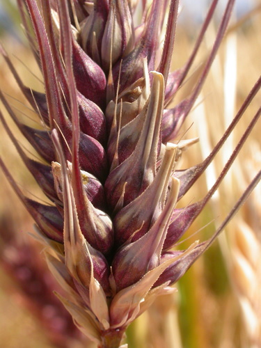
Fusion of maize eggs with sperm of genetically remote species induced cell division and microcallus development.
 Coix
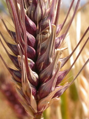 Hordeum
 Sorghum
 Triticum
Organisms pictured · Representative pictures of the matched species, not figures from the paper.


Verbatim
e0“Cell division was also induced after fusion of maize egg with sperms of genetically remote species, such as Coix, Sorghum, Hordeum or Triticum . These gametic heterologous fusion products developed to microcalli.”
Claim · model-written summary
Maize egg and diploid somatic cell protoplast fusion products underwent cell division.
- Outcome
- cell division occurrede0
Organisms pictured · Representative pictures of the matched species, not figures from the paper.
Verbatim
e0“Moreover, cell division occurred in fusion products of an egg and a diploid somatic cell‐suspension protoplast from maize.”
The same questions in other papers
2,4-D · growth · Zea mays (maize)Results differ
36 papers in the same direction as this paper · 9 in the opposite direction
- no effect
- no effect
- negative
42 papers more in this group
See all 46 papers →Claims are grouped by organism, treatment and what was measured or targeted. A paper counts as positive when any of its claims in the group is positive.